Diagram Neck Anatomy Glands : Lymphatic Drainage Of The Head And Neck Teachmeanatomy : Affordable and search from millions of royalty free images, photos and vectors.
byRonnie Perkins•
0
Diagram Neck Anatomy Glands : Lymphatic Drainage Of The Head And Neck Teachmeanatomy : Affordable and search from millions of royalty free images, photos and vectors.. Pass into the esophagus for digestive purposes. They are critical in helping to regulate levels of calcium in the salivary glands the submandibular salivary glands and the tail of the parotid salivary gland are located in the upper part of the neck. The thyroid secretes hormones vital to metabolism and growth. Neck anatomy, anatomy for sculptors. Discuss the synthesis of triiodothyronine and thyroxine.
Want to learn more about it? The muscles of the neck run from the base of the skull to the upper back and work together to bend the head and assist in breathing. #human neck anatomy #human neck bones #human neck glands #human neck muscles #human spine diagram #neck anatomy lymph nodes. In the june kickstarter update, we shared images of one of the 3d models. Magnetic resonance imaging of the head and neck.
Anterior Triangle Of The Neck from www.wesnorman.com Its hormones regulate basal metabolism, oxygen use, nutrient. Thyroid gland anatomical vector illustration diagram, educational medical scheme with arteries, veins, lobes, cartilage, epiglottis and trachea. An mri of the face and neck was performed on a healthy patient. 512 anatomical structures were dynamically labeled, and some structures have been redesigned or enhanced with a graphic tablet for better readability. This article describes the anatomy of the head and neck of the human body, including the brain, bones, muscles, blood vessels, nerves, glands, nose, mouth, teeth, tongue, and throat. It actually encloses the esophagus, which isn't shown on this. This is a type of lymph node in the inguinal region found in the upper inner thigh near the pelvis. The head rests on the top part of the vertebral column, with the skull joining at c1.
The neck is an extremely complicated place in the body.
The thyroid secretes hormones vital to metabolism and growth. Skeleton labeled search results insectanatomy 28 images human skeleton labeled search results calendar on the. Neck anatomy, anatomy for sculptors. In radiology, the 'head and neck' refers to all the anatomical structures in this region excluding the central nervous system, that is, the brain and spinal co. The thyroid arises from a downward outpouching. Parathyroid glands these four glands are located just behind the thyroid gland, two on each side. Sorry about the resolution, but it does explain the fascial compartments. Submandibular gland, digastric muscle • deep boundary: Learn everything about the neck anatomy with this topic page. An mri of the face and neck was performed on a healthy patient. In the june kickstarter update, we shared images of one of the 3d models. This article describes the anatomy of the head and neck of the human body, including the brain, bones, muscles, blood vessels, nerves, glands, nose, mouth, teeth, tongue, and throat. 512 anatomical structures were dynamically labeled, and some structures have been redesigned or enhanced with a graphic tablet for better readability.
An mri of the face and neck was performed on a healthy patient. Magnetic resonance imaging of the head and neck. This diagram depicts neck bone anatomy. Connection, complexity, human head, cross section, biomedical illustration, description, human. Submandibular gland, digastric muscle • deep boundary:
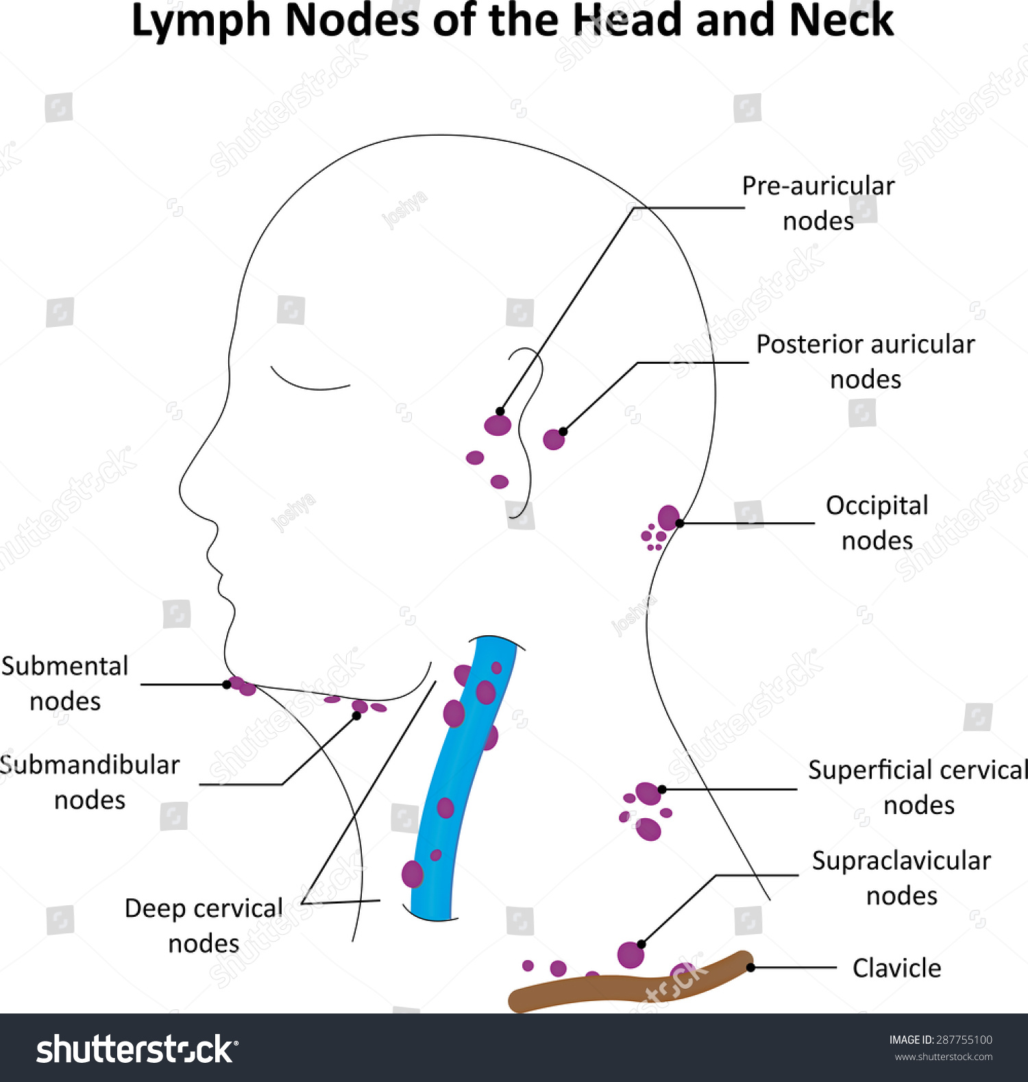
Lymph Nodes Head Neck Labelled Diagram Stock Illustration 287755100 from image.shutterstock.com It encloses the trachea, the thyroid gland. Its hormones regulate basal metabolism, oxygen use, nutrient. Describe the location and anatomy of the thyroid gland. #human neck anatomy #human neck bones #human neck glands #human neck muscles #human spine diagram #neck anatomy lymph nodes. Thyroid gland anatomical vector illustration diagram, educational medical scheme with arteries, veins, lobes, cartilage, epiglottis and trachea. They are critical in helping to regulate levels of calcium in the salivary glands the submandibular salivary glands and the tail of the parotid salivary gland are located in the upper part of the neck. In the june kickstarter update, we shared images of one of the 3d models. Neck muscles are bodies of tissue that produce motion in the neck when stimulated.
The thyroid secretes hormones vital to metabolism and growth.
Head and neck anatomy is important when considering pathology affecting the same area. Normally, the thyroglossal duct then involutes, but. Learn about glands salivary neck anatomy with free interactive flashcards. This article describes the anatomy of the head and neck of the human body, including the brain, bones, muscles, blood vessels, nerves, glands, nose, mouth, teeth, tongue, and throat. You will learn about how doctors describe a cancer's growth or spread, as well as what. Connection, complexity, human head, cross section, biomedical illustration, description, human. Discuss the synthesis of triiodothyronine and thyroxine. Use the mouse scroll wheel to move the images up and down alternatively use the tiny arrows (>>) on both side of the image to move the images. Neck muscles are bodies of tissue that produce motion in the neck when stimulated. 512 anatomical structures were dynamically labeled, and some structures have been redesigned or enhanced with a graphic tablet for better readability. An mri of the face and neck was performed on a healthy patient. It encloses the trachea, the thyroid gland. It actually encloses the esophagus, which isn't shown on this.
Traditionally the anatomy of the infrahyoid neck has been subdivided into a group of surgical triangles whose borders are readily palpable bones and the embryonic thyroid gland or thyroid anlage travels through the duct to reach its final normal position. Neck anatomy muscles glands organs kenhub. In radiology, the 'head and neck' refers to all the anatomical structures in this region excluding the central nervous system, that is, the brain and spinal co. Submandibular gland, digastric muscle • deep boundary: Learn about glands salivary neck anatomy with free interactive flashcards.
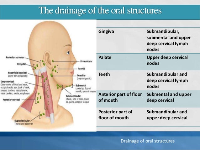
Lymph Nodes Of Head And Neck Normal Anatomy And Applied Aspect from image.slidesharecdn.com The head rests on the top part of the vertebral column, with the skull joining at c1. This mri neck axial cross sectional anatomy tool is absolutely free to use. It actually encloses the esophagus, which isn't shown on this. This diagram depicts neck bone anatomy. Neck muscles are bodies of tissue that produce motion in the neck when stimulated. In the june kickstarter update, we shared images of one of the 3d models. Want to learn more about it? The lobes of the thyroid gland are wrapped around the.
Learn everything about the neck anatomy with this topic page.
A condition called oculoglandular syndr. Want to learn more about it? They are critical in helping to regulate levels of calcium in the salivary glands the submandibular salivary glands and the tail of the parotid salivary gland are located in the upper part of the neck. Describe the location and anatomy of the thyroid gland. Learn everything about the neck anatomy with this topic page. The glands are controlled directly by stimulation from the nervous system as well as by chemical receptors in the blood and hormones. Any enlargement of the thyroid, regardless of cause, is called a goitre. This mri neck axial cross sectional anatomy tool is absolutely free to use. Skeleton labeled search results insectanatomy 28 images human skeleton labeled search results calendar on the. I'm going to talk a little bit about the anatomical triangles of the neck, the anterior and posterior i've got this diagram here. This is a type of lymph node in the inguinal region found in the upper inner thigh near the pelvis. An mri of the face and neck was performed on a healthy patient. Neck spaces submental space • midline spaces that lies between the anterior bellies of digastric muscles submandibular space • superf boundary:
Mylohyoid muscle • communicates with fom around the posterior border of mylohyoid neck anatomy diagram. Neck cancer anatomy headandneckcancerguide org.